 EN
EN  DE
DE  ES
ES  FR
FR 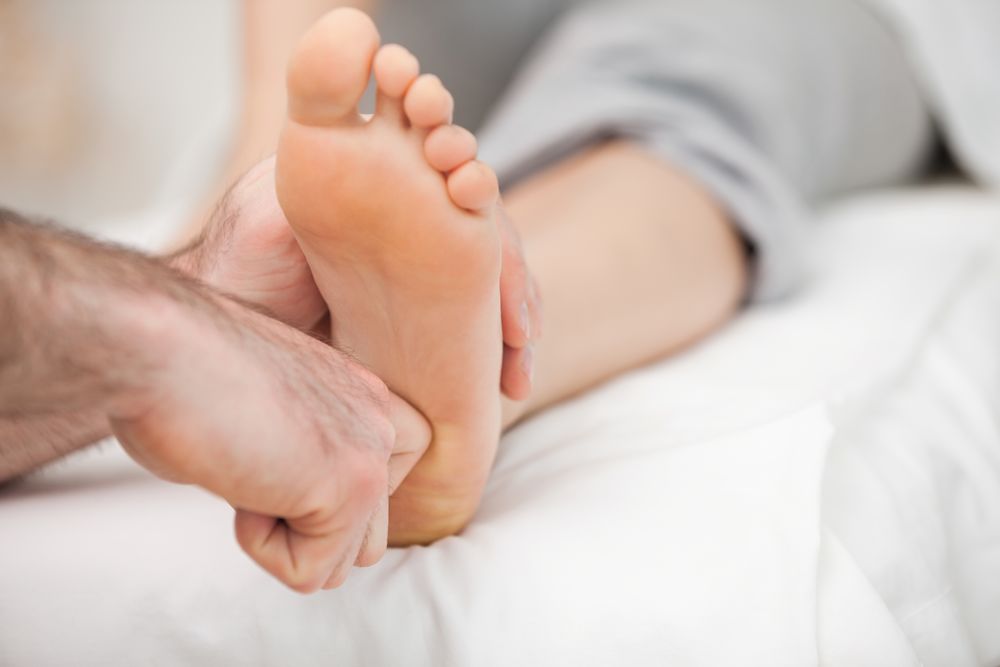
The role of osteopathy is to re-establish functional and harmonious body coordination as a whole in cases of a reversible local or global dysfunction in patients of all ages. FHL often warrants an osteopathic approach, given the multiple different level localisation of symptoms.
Locally, the tenodesis effect of the flexor hallucis longus causes blockage at the level of the subtalar joint, which has to be lifted by the “vacuum cleaner cord manoeuvre” in order for the tendon to glide freely. The subtalar blockage is often associated with blockage of the superior tibiofibular joint, through the internal tibial pronation-rotation synchronization, dysfunction of the inferior tibiofibular joint, which are often a sequel to ankle sprain and finally navicular dysfunction.
Subtalar joint mobilisation can be performed in various manners which are well documented:
- classic manoeuvre
- supine/prone positioning
- manipulation of the superior tibiofibular joint before or after the subtalar joint mobilisation depending on the case
General,
The dysfunction related to the FHL pathology consists of increased flexion in the sagittal plane of the hip and knee joints and anterior projection of the torso. Dananberg has presented the problem eloquently in his article: « Gait style as an etiology to chronic postural pain » (DANANBERG HJ: Gait style as an etiology to chronic postural pain: part 1. Functional hallux limitus. JAPMA 83:433, 1993. DANANBERG HJ: Gait style as an etiology to chronic postural pain: part 2. Postural compensatory process. JAPMA 83: 615, 1993).
Concerning the pelvis
The anterior pelvic tilt alters the muscular vector of the gluteus medius, transposing it anteriorly and in that manner engaging the piriformis in the stabilisation of the pelvis during unipedal stance. The piriformis muscle cannot adequately adapt to this situation and because of its excessive use it gradually develops a contracture. This in turn is followed by muscle shortening which limits the range of motion of the sacroiliac joint. This joint then becomes vulnerable and can end up in a fixed anterior or posterior malposition. In association to the sacroiliac joint dysfunction, a compensatory blockage of the sacrum in flexion can be observed, contralaterally to the contracted piriformis muscle. This double dysfunction can lead to rotation of the 4th and/or 5th lumbar vertebra, a frequent source of lower back pain. In that case, lower extremity length discrepancy will be observed where the “falsely” shorter leg will be ipsilateral to a posterior sacroiliac dysfunction and contralateral to an anterior sacroiliac dysfunction.
In conclusion:
anterior-superior dysfunction: seemingly longer leg ipsilaterally
posterior-inferior dysfunction: seemingly shorter leg ipsilaterally
Concerning the spine
Lumbar spine: the anterior pelvic tilt and the increased flexion at the hip joint cause a forward tilting of the torso. This tilting results in excessive overload of the posterior muscles (erector spinae) during their effort to maintain balance by counteracting the anteriorly dropped torso. The pelvic dysfunction leads to an aggravated lumbar lordosis and rigidity due to iliopsoas contracture. In that setting of articular constraint and muscular distortion, the dorsolumbar hinge finds itself prone to blockage. Blockage in rotation is especially favoured by unilateral presence of flexor hallucis longus dysfunction or strong laterality (predominant use of right or left extremity).
During clinical examination, this blockage presents as a localised contracture (trigger point), as well as limited rotational capacity of the torso towards the right or left depending on the laterality of the dysfunction. Note: in the presence of extreme kyphosis, the extension range of the dorsolumbar hinge is frequently limited.
The osteopathic approach for the release of these blockages consists of:
- use of the Fryette technique when the dysfunction is in rotation
- use of rolling movements when the dysfunction is in flexion
Cervical spine: the dysfunction can reflect on a higher lever and lead to contracture and blockage on the cervical spine. Lower extremity length discrepancy causes a lateral flexion on the frontal plane. This is compensated by scoliotic deformation of the spine. The body seeks to maintain a horizontal eye setting at the last compensatory level (C2). It is at that level that symptoms are localised in the form of headaches and rotatory limitation of the cervical spine.
Osteopathy in the pediatric setting
FHL can be detected early on in life by performing the stretch test even in infants. Later on, with the acquisition of walking skills, gait with an internal angulation of the step is frequently associated with the presence of FHL and pelvic dysfunction related to the numerous falls on the glutes during the process. Manipulation of the hind foot and daily stretching exercises of the long flexor muscle performed by the parents are very effective in restoring balance and a more harmonious gait of the child. It is appropriate to follow up the child and ensure the free excursion of the flexor hallucis longus and the mobility of the subtalar joint.
In case of family history of hallux valgus, an early treatment for FHL can probably prevent or reduce the risk of that deformity. Young ice skaters or dancers are particularly exposed to the risk of fore foot deformation, since training in demi-pointe position is a predisposing factor for hypertrophy of the flexor hallucis longus muscle which in turn predisposes for functional hallux limitus.
Summary
Osteopathic treatment is complementary to physical therapy and if well applied, can improve significantly patients’ gait and balance from a very early age.